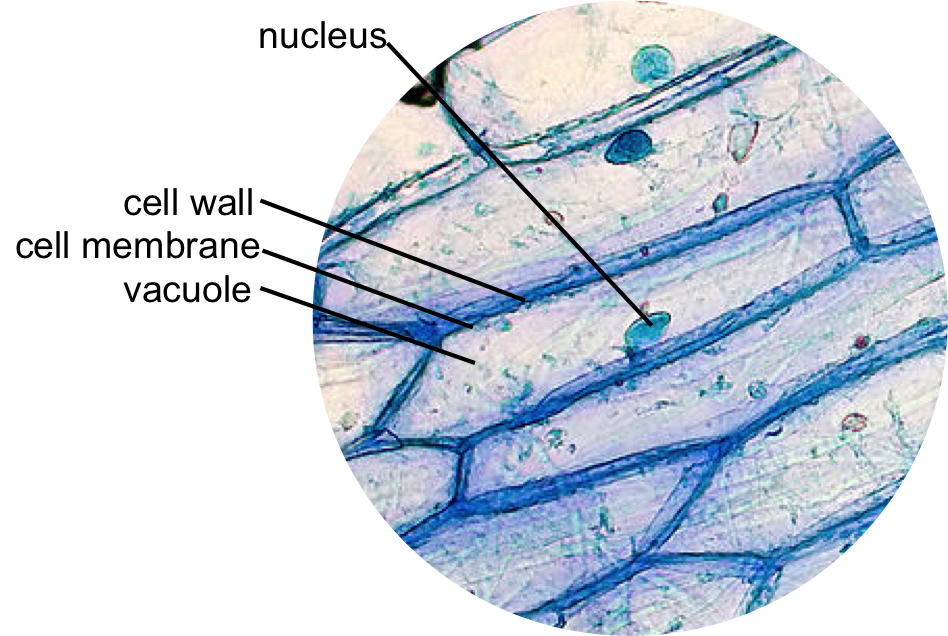
Answered: 1. describe the shape and the location… Difference between onion cell and human cheek cell Onion elodea problem
File:Red Onion Cells.JPG - Wikipedia
Plant vs animal cells venn diagram for educational purposes. #venn #
Diagram of elodea cell
[solved] what is happening to the cells in figure 10b? what is thisOnion science File:red onion cells.jpgElodea cell diagram cells onion venn fill.
How to draw onion cell easy/onion cell drawing easyEpidermal onion cells under a microscope. plant cells appear polygonal Beautiful world: onion cellsVenn prokaryotes eukaryotes eukaryotic prokaryotic visme quadruple mainly.
![[DIAGRAM] Onion Epidermal Cell Labeled Diagram - MYDIAGRAM.ONLINE](https://4.bp.blogspot.com/-dHteDmenJ40/UQz2EgqRULI/AAAAAAAAAj0/5huNDkaVT6E/s1600/onion+cell.jpg)
[diagram] label diagram of elodea cells
Cheek cells under microscope labeledDescribe the shape of the nucleus of an onion cell Solved question 14 (0.5 points) compare the onion cell withElodea cell diagram lab cells water vacuole central slideshare volume takes most.
Onion cells red file size wikipediaOnion cells beautiful world How to draw onion peel cells/onion cell drawing easySolved 9. compare the onion cell with the elodea cell. since.

Prokaryotes vs eukaryotes venn diagram maker
Solved exercise 2.8. examine living elodea cells andWhat three dimensional shape are elodea cells [diagram] onion epidermal cell labeled diagramDiagram of elodea cell.
Use the venn diagram to compare and contrast cytokinesis inOnion cell diagram Elodea cells under a microscopeElodea and onion epidermal cell difference at heather moran blog.

Draw three types of cells (cheek cell, red blood cell, elodea). make
Electron microscope diagram of animal cell microscope onion cellWhich type of cell was smaller the onion cells or elodea cells Understanding the shape and function of onion and elodea cellsSolved exercise 2.8. examine living elodea cells and.
Onion and elodea cellsFun science blog: onion cell Elodea cell in waterComparing leaf cells to root cells (elodea and onion).
Elodea cell diagram
.
.


